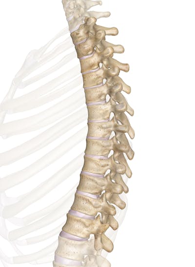
Thoracic Vertebrae : Spine Back Bones Thoracic Vertebrae Ranzcrpart1 Wiki Fandom - Thoracic vertebrae are unique among the bones of the spine in that they are the only vertebrae that support ribs and have overlapping spinous processes.
Get link
Facebook
X
Pinterest
Email
Other Apps
Thoracic Vertebrae : Spine Back Bones Thoracic Vertebrae Ranzcrpart1 Wiki Fandom - Thoracic vertebrae are unique among the bones of the spine in that they are the only vertebrae that support ribs and have overlapping spinous processes.. Clinical presentations of shoulder pain online course: Types of thoracic vertebrae injury and. Learn about thoracic vertebra with free interactive flashcards. The cervical vertebrae, the thoracic. Thoracic facet joint disorder is a common thoracic disorder in clinic, inducing pain and discomfort at the dislocated thoracic vertebrae, radiating to pain of the neck and back.
Learn about thoracic vertebra with free interactive flashcards. Types of thoracic vertebrae injury and. The thoracic vertebrae are bones located between the cervical and lumbar vertebrae. 6 different views demonstrated with the rotation step of 45°. The top view of the 4th thoracic vertebra.
Thoracic Vertebrae T2 T8 from www.getbodysmart.com The thoracic spine is comprised of 12 vertebrae labeled t1 through t12. See more ideas about thoracic vertebrae, thoracic, vertebrae. Схема поясничного сплетения и его нервов (иванов г.ф., 1949). Vertebrae thoracicae) comprise the middle portion of the vertebral column and are characterized by their articulation with ribs. Clinical presentations of shoulder pain explore how. Bony arches project from the back of each vertebral body forming a hollow protective space containing the spinal cord. Choose from 500 different sets of flashcards about thoracic vertebra on quizlet. Vertebral body is heart shaped.
How many thoracic vertebrae are there?
Clinical presentations of shoulder pain explore how. The thoracic vertebrae are located in the thorax posterior and medial to the ribs. Types of thoracic vertebrae injury and. What do you prefer to learn with? For a basic anatomic description of the structure of. Related online courses on physioplus. Choose from 500 different sets of flashcards about thoracic vertebra on quizlet. Thoracic vertebrae blood supply and lymphatics The cervical vertebrae, the thoracic. There are 12 thoracic vertebrae in humans, and these bones increase in size as you move down the body. Above are the cervical vertebrae and below are the lumbar, sacral, and coccygeal vertebrae. Learn about thoracic vertebrae anatomy & function. The body resembles that of a cervical vertebra.
The top view of the 4th thoracic vertebra. The twelve thoracic vertebrae make up the middle portion of the vertebral column. Choose from 500 different sets of flashcards about thoracic vertebra on quizlet. Above are the cervical vertebrae and below are the lumbar, sacral, and coccygeal vertebrae. In human, thoracic vertebrae consists of 12 bones.
Thoracic Vertebrae from innerbody.imgix.net From top to down, t1, t2, …, t12. Its upper surface is lipped laterally and beveled anteriorly. Thoracic facet joint disorder is a common thoracic disorder in clinic, inducing pain and discomfort at the dislocated thoracic vertebrae, radiating to pain of the neck and back. Clinical presentations of shoulder pain explore how. Bony arches project from the back of each vertebral body forming a hollow protective space containing the spinal cord. The thoracic vertebrae are located in the thorax posterior and medial to the ribs. Learn about thoracic vertebra with free interactive flashcards. The thoracic vertebrae have four features which distinguish them from other vertebrae:
The cervical vertebrae, the thoracic.
The top thoracic vertebra, t1, connects with c7 in the cervical spine above while the bottom thoracic vertebra, t12, connects. Vertebrae thoracicae) comprise the middle portion of the vertebral column and are characterized by their articulation with ribs. The thoracic spine is comprised of 12 vertebrae labeled t1 through t12. The thoracic vertebrae have four features which distinguish them from other vertebrae: The body resembles that of a cervical vertebra. They form the region of the spinal column inferior to the cervical vertebrae of the neck and superior to the lumbar. Learn about thoracic vertebra with free interactive flashcards. In total there are 12 thoracic vertebrae. The twelve thoracic vertebrae make up the middle portion of the vertebral column. The thoracic vertebrae each articulate with two costal bones, otherwise known as ribs, to form the major structural elements of the thoracic cavity—also known as the thorax or simply the chest. The thoracic vertebrae possess slightly triangular bodies with flat superior and inferior end plates and longer pedicles than the cervical vertebrae that cover the intervertebral foraminae. The top view of the 4th thoracic vertebra. Thoracic vertebrae blood supply and lymphatics
The top thoracic vertebra, t1, connects with c7 in the cervical spine above while the bottom thoracic vertebra, t12, connects. The thoracic vertebrae are bones located between the cervical and lumbar vertebrae. From top to down, t1, t2, …, t12. See more ideas about thoracic vertebrae, thoracic, vertebrae. 6 different views demonstrated with the rotation step of 45°.
Thoracic Vertebrae T1 T12 Anatomy Youtube from i.ytimg.com Related online courses on physioplus. It is broad and not heart shaped. The thoracic vertebrae have four features which distinguish them from other vertebrae: The presence of costal facet/facets on the sides of their bodies for articulation with the heads of the ribs is how they can be identified or detected. Position of the thoracic vertebrae (shown in red). The body resembles that of a cervical vertebra. What do you prefer to learn with? The top thoracic vertebra, t1, connects with c7 in the cervical spine above while the bottom thoracic vertebra, t12, connects.
In the eleventh thoracic vertebra the body approaches in its form and size to that of the lumbar vertebræ.
Above are the cervical vertebrae and below are the lumbar, sacral, and coccygeal vertebrae. The thoracic vertebrae are bones located between the cervical and lumbar vertebrae. The twelve thoracic vertebrae make up the middle portion of the vertebral column. Bony arches project from the back of each vertebral body forming a hollow protective space containing the spinal cord. Given the twelve thoracic vertebrae are largely similar, most are considered typical thoracic vertebrae with the exceptions t1 and t9 to t12. For a basic anatomic description of the structure of. Vertebrae thoracicae) comprise the middle portion of the vertebral column and are characterized by their articulation with ribs. What do you prefer to learn with? The thoracic vertebrae are located in the thorax posterior and medial to the ribs. The body resembles that of a cervical vertebra. Types of thoracic vertebrae injury and. Like most other spinal vertebrae, the thoracic vertebral bodies are rounded. See more ideas about thoracic vertebrae, thoracic, vertebrae.
The thoracic vertebrae are bones located between the cervical and lumbar vertebrae thor. The presence of costal facet/facets on the sides of their bodies for articulation with the heads of the ribs is how they can be identified or detected.


Comments
Post a Comment